News

COVID-19.info

Popularization of research advances on COVID-19

Website developed by 100pour100 MEDECINE

The plasticity of the spike protein

Over the last two decades, three coronaviruses have posed a danger to public health: SARS-CoV-1 in 2002-2003; MERS-CoV in 2012 and finally SARS- CoV- 2 in 2020. Coronaviruses are “enveloped” viruses, meaning that they are covered by an external phospholipid layer whose composition is close to the membranes of human cells. The spike (S) protein is threaded into this envelope and oriented outwards to recognize the ACE2 receptor. This interaction enables the virus to enter cells, through the fusion of the viral envelope and the membrane of the infected cell.

The spike is organized in homotrimere (3 identical S proteins are linked together) that form large protuberances on the surface of the virus. Its distal domain (the furthest away) is made up of S1s which attach to the receptor via the RBD (Receptor Binding Domain). The S2 proximal domain (nearest to the envelope) governs the process of fusion via the fusion peptide (FP), a hydrophobic region that slips into the cellular membrane. SARS- CoV- 1 and SARS-CoV-2 have S proteins that are identical at 76% and attach to the same ACE2 receptor. However, SARS-CoV-2 is more infectious and the D416G mutation that is currently circulating increases infectiousness for reasons that we don’t fully understand.

We can schematically represent Spike as follows:

Researchers at the Shanghai Academy of Sciences studied the architectural composition of the S protein with the help of electronic cryomicroscopy, with a resolution of 2,7/3,8 Å. By comparing in detail the different states in which S exists, they reconstituted the successive changes in the structure which take place during infection, as follows:

  • Within the “free” virus, S exists at 94% in a “closed” (pre-fusion) form: the FPs are wrapped up and inactive, and the RBD are masked. A small fraction of S is transiently open (6%), with a single RBD (out of 3, since S is trimeric) on the surface to allow fixation to ACE2. This shape differs from the previous one by the rotation and downward movement of S1.
  • Fixing to ACE2 shifts the energy balance towards a majority of “open” forms with RBD exposed. The ACE2-RBD link up then causes changes in equilibrium on the surface, modifying the overall structure of S1.
  • These structural changes cause the underlying S2 region to be reorganized, exposing the FP towards the exterior and activating membrane fusion.

In addition to this chronological sequence, these structural elements provide details relating to the RBD-ACE2 fixation zones. Moreover, they indicate that the S protein is extremely sensitive to ACE2: its fixing to the receptor changes its state from 94% closed to 100% open. In SARS-CoV-1, 72,4% of S is already open and the presence of ACE2 increases this figure to 76%. This may explain why SARS-CoV-2 is more infectious

This can be schematically represented as follows:

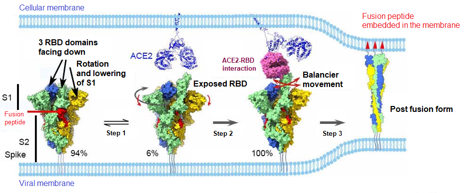
What’s more, the authors show that the amino acid D614 interacts with the FP and contributes enormously to keeping the structure closed. In the D614G mutation, the amino acid aspartate (D) is mutated into glycine (G), which has no loaded side chain. This will therefore reduce interactions across the structure, allowing the FP to partially activate. The D614G mutation is more infectious because it makes S even more sensitive to ACE2.

The authors also suggest that in SARS-CoV-2, the spike’s mostly closed conformation is an escape mechanism for neutralizing antibodies (as with HIV), which mainly target RBD. In comparison, SARS-CoV-1 and MERS-CoV are more sensitive to these antibodies since they only have 27,6% and 5,4% respectively of the S closed. This work provides very important information for the development of vaccines, since the S protein is the major target of antibodies when the immune system is triggered.

error: Content is protected !!