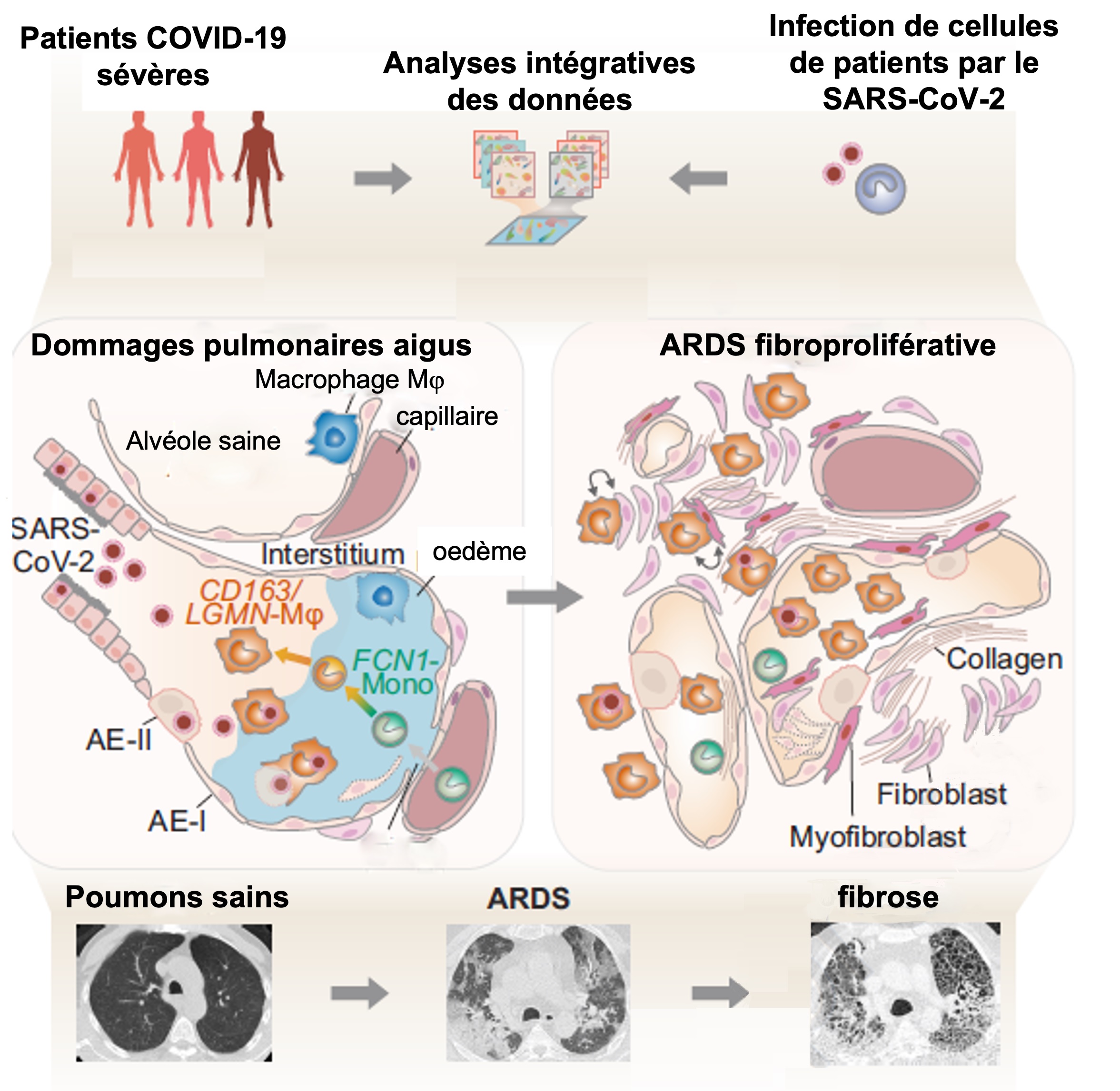
Around 5% of COVID-19 patients develop Acute Respiratory Distress Syndrome (ARDS), the mechanisms of which are still poorly understood. The development of non- COVID-19 cases of ARDS has 3 stages: damage to the alveolar barrier and the appearance of pulmonary edema, proliferation of epithelial cells to restore this barrier and, in some patients, a phase of idiopathic pulmonary fibrosis (IPF) associated with respiratory distress and high mortality. These stages are regulated by different subtypes of macrophages.
Severe cases of COVID-19 are linked to hyper-inflammation where monocytes and macrophages are the major players. German researchers at the University of Charité-Berlin have studied their role in the ARDS of COVID-19. To do so, they isolated and analysed, using several integrative approaches (genetics, proteomics and imaging), cells from the bronchoalveolar fluids of 2 cohorts of severely ill COVID-19 patients with ARDS.
Tissue analysis showed significant alveolar damage, fibroproliferative responses, and signs of pneumonia. A marked enrichment of monocytes and macrophages (in particular CD163+) was noted). RNA of SARS-CoV-2 was detected, principally in these cells. Six different populations were identified, all specifically linked to tissue damage and their repair. During the first stages of ARDS, some of these populations dominate in the alveolar chambers (FCN-1-Monocytes, Mono-Mᵠ, CD163/LGMN-Mᵠ), then are progressively replaced by others in the advanced stages (AMᵠ1, AMᵠ2).
At the genetic level, these researchers showed that the CD163/LGMN-Mᵠ have a profile that is similar to that of macrophages at the origin of FPI (the genes SPP1, TGFB1, LGMN, CCL18). During the development of ARDS, these macrophages interacted more and more with lung fibroblasts, a phenomenom which is very marked in fatal cases of COVID-19. These macrophages were localised in the same areas as the collagen produced by the fibroblasts, which accumulates, the whole constituting an environment favourable to the development of fibrosis. There was progressive consolidation and cross-linking, a sign of fibroproliferative ARDS (hypertrophic scarring) where lung tissue remodels, impairing ventilation.
To conclude, the researchers infected monocytes (from healthy patients) with SARS-CoV-2 in vitro. The results showed that SARS-CoV-2, but not influenza A, induces in cells a differentiation programme identical to that in macrophages linked to FPI.
This work clarifies the pathological mechanisms of ARDS linked to severe cases of COVID-19. During the healing of lung damage linked to infection, certain subtypes of macrophages colonise the lungs. They can be directly reprogrammed by SARS-CoV-2 to a profibrotic process. In some patients, they become predominant little by little, secreting specific growth factors (TGF-ᵠ), and causing the expansion of fibroblasts and an abnormal secretion of collagen, ultimately remodelling pulmonary tissues and altering respiration. Unlike the FPI, which are progressive and irreversible, the fibroses linked to COVID-19 occur more rapidly and are partially reversible, but more lethal. These macrophages (CD163/LGMN-Mᵠ) could therefore be used as bio-markers so as to predict the development of COVID-19 ARDS and allow patients to be treated with antifibrotics.