How are they obtained ?
The distal lung environment, principally targeted by SARS-CoV-2, is composed of terminal bronchioli and alveoli where gaseous interaction with the blood takes place. In certain patients, this infection brings about acute respiratory distress syndrome, though we are not sure how.
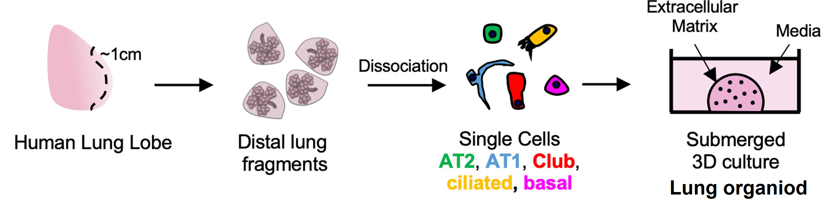
However, researchers at the University of Stanford have managed to create human pulmonary organoids by using primary stem cells derived from alveolar epithelial cells AT2 (pneumocytes) and basal KRT5+ (for maintenance of pseudostratified epithelium). The AT2 pneumocytes renew themselves efficiently and differentiate into AT1 cells to mimic the epithelium lining the alveoli. The basal cells differentiate into hair cells and bronchiole cells. The whole reorganises itself to form a complex polarised environment with microvilli and apical junctions which imitate the exterior lung surface. These organoids therefore provide a very satisfactory replica of the natural lung environment.
How do these organoids react to the virus?
Around 10% of the basal and epithelial organoid cells are infected by SARS-CoV-2 due to the ACE2 receptor apical expression, just as in natural lungs. This system has also shown infection by the H1N1 flu virus to be inhibited by the nucleoside analogue Fdc.
However, and unexpectedly, the bronchiole cells could be a new target for the virus, damaging the production of the protective lining of lung tissues (glycosaminoglycans) and thereby stimulating the infection cycle.
These organoids are therefore suitable for use in research on antiviral compounds, even if their bronchiolotic compounds overreact. This new model, viable over the long term and simple to cultivate, could also allow study of other infectious lung diseases, interstitial or neoplastic, as well as finding application in precision medicine.